Our Technology
Utilizing Cutting-Edge Technologies to Evaluate Eye Health

Optomap
The Optomap is a camera that uses a wide angle Nikon lens to see 90% of the inside of the eye in one image. Its purpose is to help patients better understand their own eye health by allowing them to see their eyes in a different way. These images are digital and can be referred back to for evaluation of changes inside of the eye. This makes exams more comfortable and quicker for patients. Depending on the doctors findings patients may not need to be dilated every year as was previously needed.

Optical Coherence Tomography (OCT)
The optical coherence tomography (OCT) is a medical instrument used by our office to obtain high-resolution images of the retina at the back of the eye. It works by measuring light entering and leaving your eye. These measurements are used to construct a detailed, layered map of the retina, which can help diagnose eye conditions such as macular degeneration, glaucoma, and diabetic retinopathy.

Virtual Field
This device uses the power of virtual reality to make a previously uncomfortable test much more convenient. It will measure your peripheral vision and help determine any blind spots you have. This is essential in the diagnosis of glaucoma, strokes and other conditions which can affect peripheral vision. It is even portable so if a patient has limited mobility we can still bring it to them to evaluate their vision.

iCare Tonometer
Measuring the pressure of your eye is an important part of your eye exam as having pressures too low or too high can indicate a problem. Many people dread the “air puff test”, but with the iCare tonometer you no longer have to worry. This portable, handheld device quickly and accurately measures eye pressure with no discomfort. It can even measure eye pressure without removing your contact lenses.
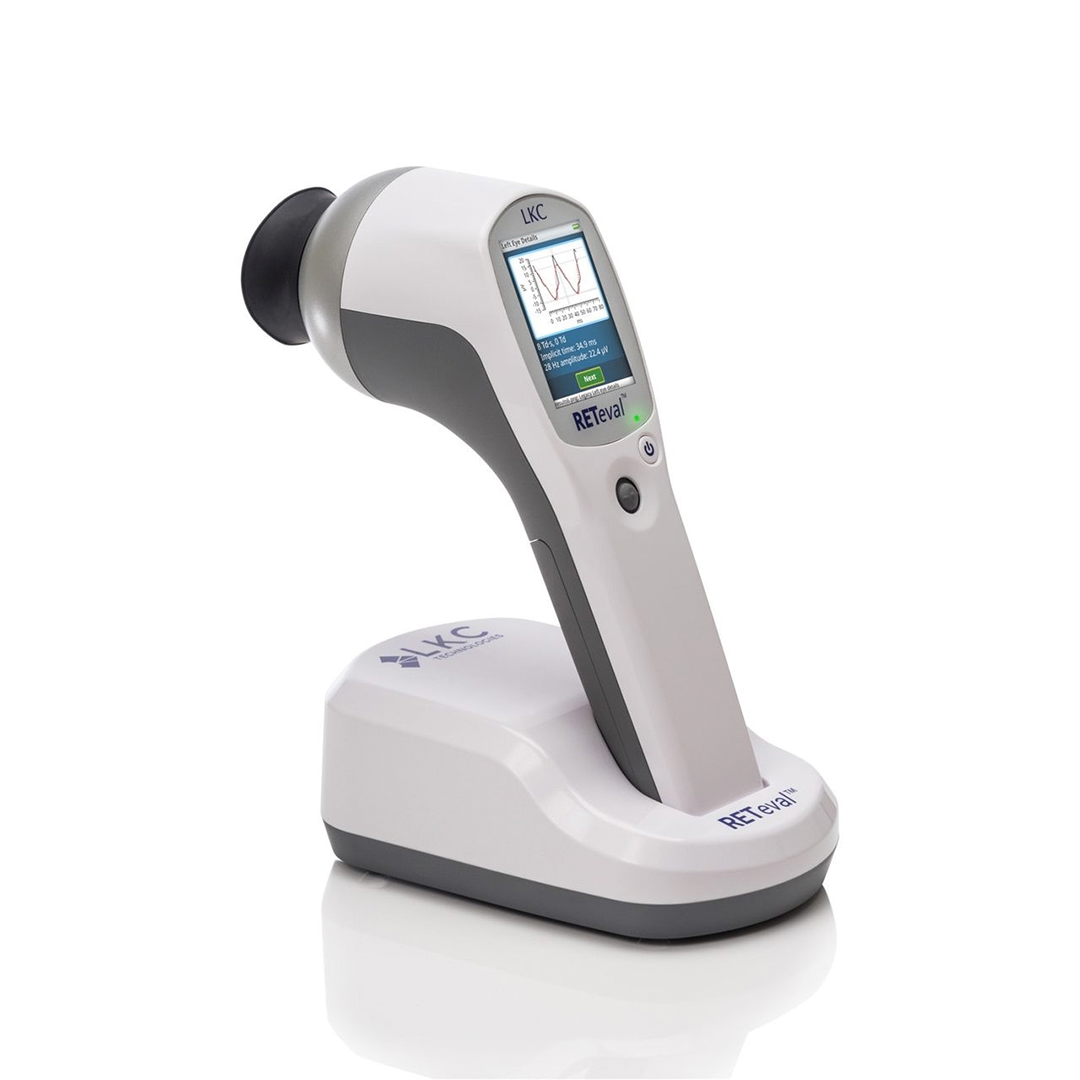
RETeval ERG
The RETeval ERG is a portable electroretinography (ERG) device used in eyecare to measure the electrical activity of the retina (the light-sensitive layer at the back of the eye) in response to light stimuli. It provides an objective assessment of retinal function without requiring pupil dilation or extensive preparation.

Rabin Cone Contrast Test
The Rabin Cone Contrast Test (RCCT) is a quantitative test of color vision that specifically measures the sensitivity of each of the three types of cone photoreceptors (red, green, and blue cones) in the human eye. Unlike traditional color vision tests (such as Ishihara plates), it provides an objective, numerical score for each cone type, making it highly useful in eyecare for both screening and monitoring visual function.
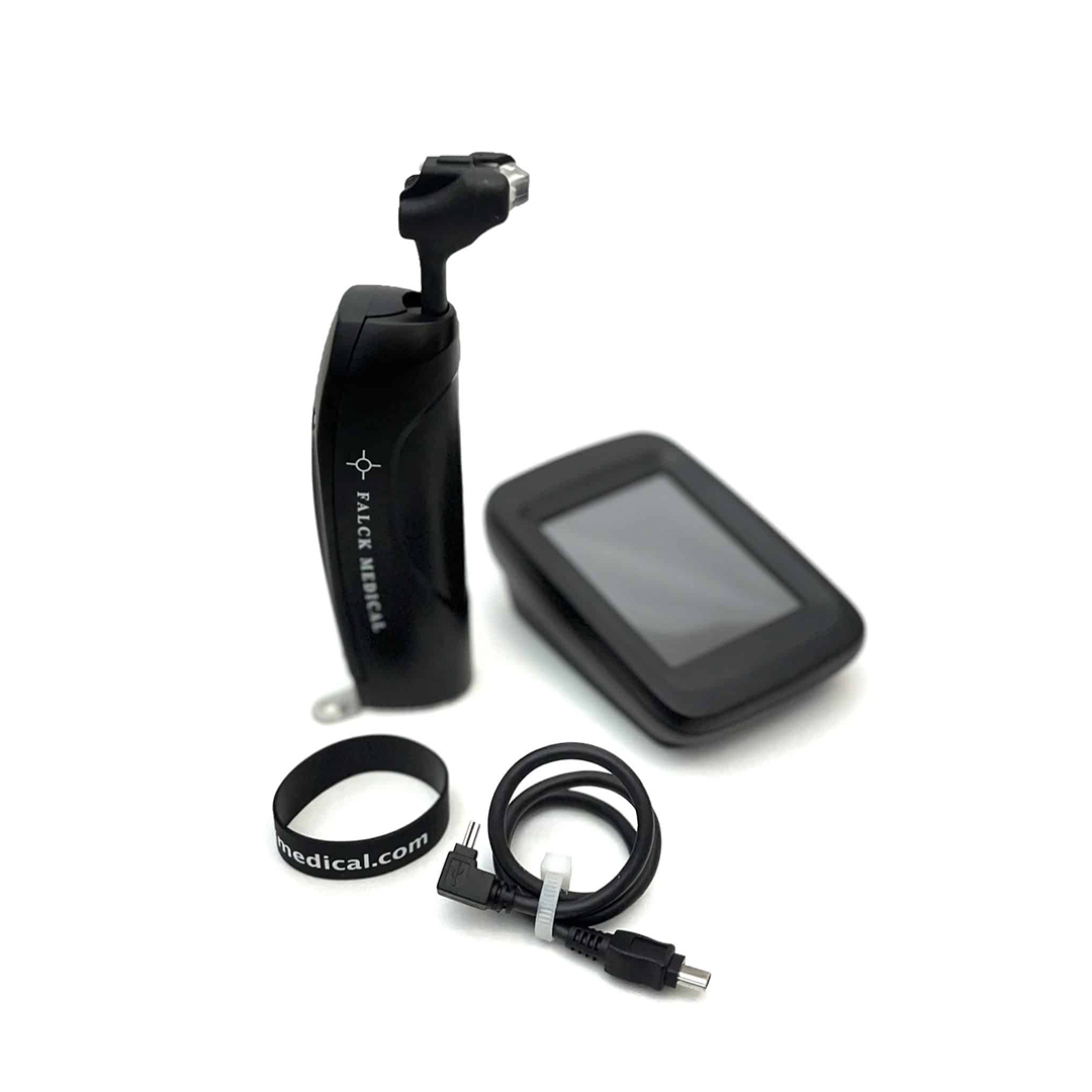
Falck Tonometry
The Falck Medical Multifunction Tonometer (FAT1) is a slit‑lamp-mounted ophthalmic diagnostic device cleared by the U.S. FDA in 2017 for multiple clinically important measurements: applanation tonometry, tonography and ophthalmodynamometry.
Transcranial Doppler (TCD)
A Transcranial Doppler (TCD) is a non-invasive ultrasound test that measures blood flow velocity in the brain’s major intracranial arteries, most often through thin areas of the skull (like the temporal bone).
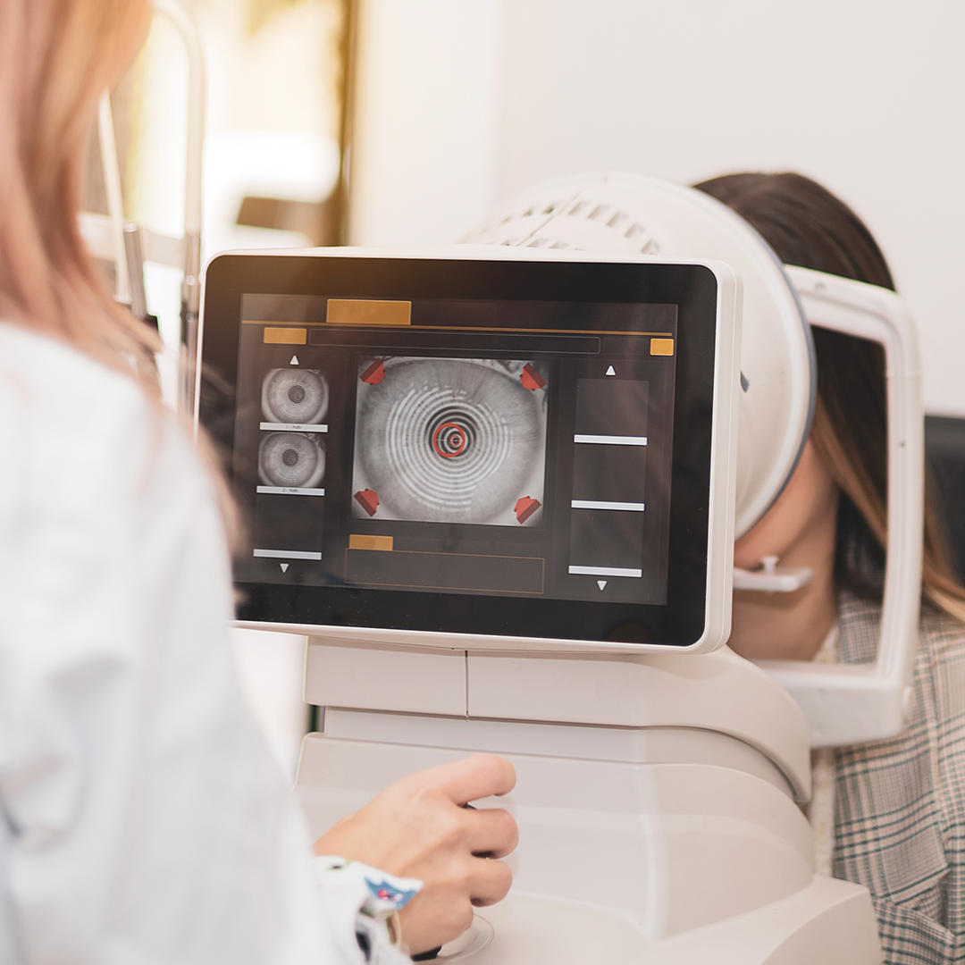
Topographer
A corneal topographer (often just called a “topographer”) is a diagnostic device that maps the curvature and shape of the cornea, the clear front surface of the eye.
It produces a color-coded map that shows steep and flat areas of the cornea.
Most systems work by projecting concentric rings (Placido disk) or grids of light onto the cornea and analyzing the reflected image.
More advanced versions (e.g., Scheimpflug imaging, OCT-based systems) also measure corneal thickness and anterior segment structures.
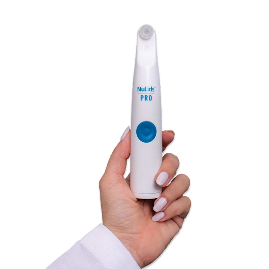
NuLids Pro
NuLids PRO is a professional-grade eyelid cleaning / hygiene device used in-office (and with a home version) to clean and stimulate the eyelid margins.
Quick Links
Helpful Articles
article_category: cosmetic
article_category: eye surgery co-management
article_category: products
article_category: vision therapy
article_category: dogs
article_category: technology
article_category: general
article_category: eye health
article_category: aesthetics
article_category: services
article_category: faqs
article_category: exotic
article_category: large animal
article_category: contact lenses
article_category: surgical procedures
article_category: health
article_category: psychiatry
article_category: conditions
article_category: restorative
article_category: preventative
article_category: cats
article_category: eyeglasses
article_category: ocular disease management
[]
Flowbite is an open-source library of interactive components built on top of Tailwind CSS including buttons, dropdowns, modals, navbars, and more.
Check out this guide to learn how to get started and start developing websites even faster with components on top of Tailwind CSS.

All Eye Care Services
Find personalized eye care excellence with our comprehensive services. From eye exams to advanced diagnostics, trust us for all your vision needs.

Keep In Touch
For non-urgent questions or to learn more about our services, contact us today!
Quick Links
Our Services
Contact Info
- 57 North St, Ste 103
Danbury, CT 06810 - Phone: (203) 748-7393
- Fax: (203) 743-2825
Hours of Operation
Hours of Operation
- Monday 8:00am - 4:00pm
- Tuesday 8:00am - 4:00pm
- Wednesday Closed
- Thursday 8:00am - 4:00pm
- Friday 8:00am - 4:00pm
- Saturday 8:00am - 12:00pm
- Sunday Closed
- Monday
- Tuesday
- Wednesday
- Thursday
- Friday
- Saturday
- Sunday
- Monday
- Tuesday
- Wednesday
- Thursday
- Friday
- Saturday
- Sunday
© 2026 Klear Sight Eyecare Center. All rights Reserved - Accessibility Statement - Privacy Policy - Sitemap
Managed and Designed by ![]()